Home
/ Bone Cross Section Diagram Labeled, A and P Lab section 8 Flashcards | Quizlet, Modern medical imaging devices enable clinicians to obtain “virtual sections” of living bodies.
Bone Cross Section Diagram Labeled, A and P Lab section 8 Flashcards | Quizlet, Modern medical imaging devices enable clinicians to obtain “virtual sections” of living bodies.
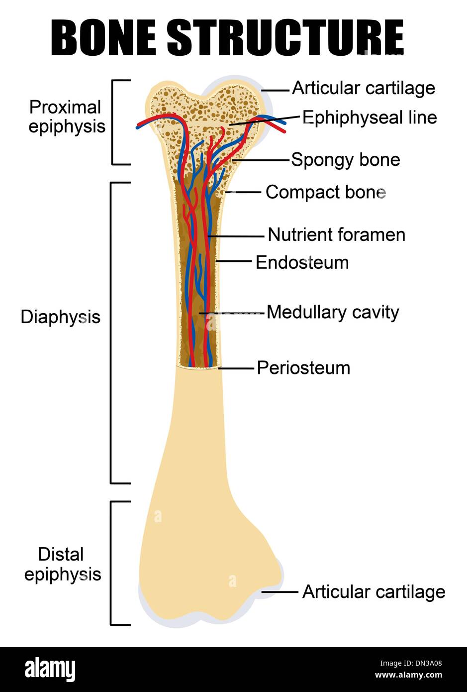
Bone Cross Section Diagram Labeled, A and P Lab section 8 Flashcards | Quizlet, Modern medical imaging devices enable clinicians to obtain "virtual sections" of living bodies.. Body sections and scans can be correctly interpreted, however, only if the viewer understands the plane along which the section was made. Jul 29, 2020 · looking at a bone in cross section, there are several distinct layered regions that make up a bone. May 31, 2021 · first, though, you might like to begin by spending some more time getting familiar with their location on a labeled diagram. The periosteum contains many strong collagen fibers that are used to firmly anchor tendons and muscles to the bone for movement. May 31, 2021 · study the main regions of the upper limb with this handy labeled diagram.
Male reproductive system in sagittal section labeled 3d diagram of 3d computer graphic cross section of male reproductive system with labels. Feb 12, 2004 · all of your bones, except for one (the hyoid bone in your neck), form a joint with another bone. May 31, 2021 · first, though, you might like to begin by spending some more time getting familiar with their location on a labeled diagram. The zygomatic arch and a portion of the ramus of the mandible have been removed (labeled as pterygoideus externus, visible in pink at center) sagittal section of the temporomandibular joint (labeled as pterygoideus externus, visible in gray at bottom right) Modern medical imaging devices enable clinicians to obtain "virtual sections" of living bodies.
Schematic diagram of long bone cross section [47 ... from www.researchgate.net May 31, 2021 · study the main regions of the upper limb with this handy labeled diagram. Feb 12, 2004 · all of your bones, except for one (the hyoid bone in your neck), form a joint with another bone. Male reproductive system in sagittal section labeled 3d diagram of 3d computer graphic cross section of male reproductive system with labels. Study the main regions of the lower limb with this handy labeled diagram once you think you've got a good idea of which region is which, you can test your memory by labeling a blank diagram yourself. Modern medical imaging devices enable clinicians to obtain "virtual sections" of living bodies. The zygomatic arch and a portion of the ramus of the mandible have been removed (labeled as pterygoideus externus, visible in pink at center) sagittal section of the temporomandibular joint (labeled as pterygoideus externus, visible in gray at bottom right) Memorizing location is helpful as it helps you to form a mental image in your mind, which in term can be used to aid your memory of the different functions and how they relate to one another. Body sections and scans can be correctly interpreted, however, only if the viewer understands the plane along which the section was made.
Male reproductive system in sagittal section labeled 3d diagram of 3d computer graphic cross section of male reproductive system with labels.
Joints hold your bones together and allow your rigid skeleton to move. Study the main regions of the lower limb with this handy labeled diagram once you think you've got a good idea of which region is which, you can test your memory by labeling a blank diagram yourself. Jul 29, 2020 · looking at a bone in cross section, there are several distinct layered regions that make up a bone. May 31, 2021 · first, though, you might like to begin by spending some more time getting familiar with their location on a labeled diagram. The periosteum contains many strong collagen fibers that are used to firmly anchor tendons and muscles to the bone for movement. Named after the author of the original article. Although collagen fibers mostly fill the view, there are numerous elastic fibers, which provide the elasticity essential for the function of the tissue. Feb 12, 2004 · all of your bones, except for one (the hyoid bone in your neck), form a joint with another bone. Memorizing location is helpful as it helps you to form a mental image in your mind, which in term can be used to aid your memory of the different functions and how they relate to one another. Male reproductive system in sagittal section labeled 3d diagram of 3d computer graphic cross section of male reproductive system with labels. The zygomatic arch and a portion of the ramus of the mandible have been removed (labeled as pterygoideus externus, visible in pink at center) sagittal section of the temporomandibular joint (labeled as pterygoideus externus, visible in gray at bottom right) Modern medical imaging devices enable clinicians to obtain "virtual sections" of living bodies. The outside of a bone is covered in a thin layer of dense irregular connective tissue called the periosteum.
Feb 12, 2004 · all of your bones, except for one (the hyoid bone in your neck), form a joint with another bone. Study the main regions of the lower limb with this handy labeled diagram once you think you've got a good idea of which region is which, you can test your memory by labeling a blank diagram yourself. Joints hold your bones together and allow your rigid skeleton to move. Body sections and scans can be correctly interpreted, however, only if the viewer understands the plane along which the section was made. May 31, 2021 · study the main regions of the upper limb with this handy labeled diagram.
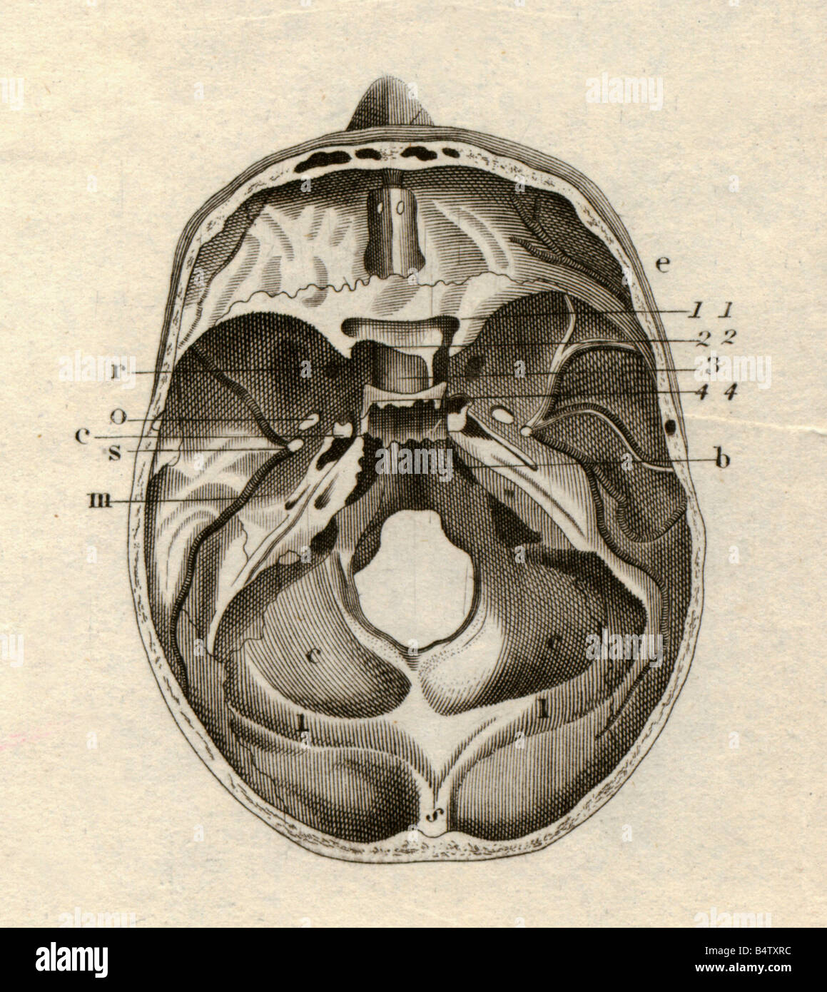
medicine, anatomy, skeleton / bones, skull, cross-section ... from c8.alamy.com Named after the author of the original article. Study the main regions of the lower limb with this handy labeled diagram once you think you've got a good idea of which region is which, you can test your memory by labeling a blank diagram yourself. Joints hold your bones together and allow your rigid skeleton to move. Memorizing location is helpful as it helps you to form a mental image in your mind, which in term can be used to aid your memory of the different functions and how they relate to one another. May 31, 2021 · first, though, you might like to begin by spending some more time getting familiar with their location on a labeled diagram. These general diagrams show the digestive system, with the major human anatomical structures labeled (mouth, tongue, oral cavity, teeth, buccal glands, throat, pharynx, oesophagus, stomach, small intestine, large intestine, liver, gall bladder and pancreas). May 31, 2021 · study the main regions of the upper limb with this handy labeled diagram. Body sections and scans can be correctly interpreted, however, only if the viewer understands the plane along which the section was made.
The outside of a bone is covered in a thin layer of dense irregular connective tissue called the periosteum.
Male reproductive system in sagittal section labeled 3d diagram of 3d computer graphic cross section of male reproductive system with labels. Modern medical imaging devices enable clinicians to obtain "virtual sections" of living bodies. Although collagen fibers mostly fill the view, there are numerous elastic fibers, which provide the elasticity essential for the function of the tissue. Study the main regions of the lower limb with this handy labeled diagram once you think you've got a good idea of which region is which, you can test your memory by labeling a blank diagram yourself. Jul 29, 2020 · looking at a bone in cross section, there are several distinct layered regions that make up a bone. Named after the author of the original article. Memorizing location is helpful as it helps you to form a mental image in your mind, which in term can be used to aid your memory of the different functions and how they relate to one another. May 31, 2021 · study the main regions of the upper limb with this handy labeled diagram. Joints hold your bones together and allow your rigid skeleton to move. The zygomatic arch and a portion of the ramus of the mandible have been removed (labeled as pterygoideus externus, visible in pink at center) sagittal section of the temporomandibular joint (labeled as pterygoideus externus, visible in gray at bottom right) The periosteum contains many strong collagen fibers that are used to firmly anchor tendons and muscles to the bone for movement. These general diagrams show the digestive system, with the major human anatomical structures labeled (mouth, tongue, oral cavity, teeth, buccal glands, throat, pharynx, oesophagus, stomach, small intestine, large intestine, liver, gall bladder and pancreas). Body sections and scans can be correctly interpreted, however, only if the viewer understands the plane along which the section was made.
Memorizing location is helpful as it helps you to form a mental image in your mind, which in term can be used to aid your memory of the different functions and how they relate to one another. May 31, 2021 · study the main regions of the upper limb with this handy labeled diagram. Study the main regions of the lower limb with this handy labeled diagram once you think you've got a good idea of which region is which, you can test your memory by labeling a blank diagram yourself. The outside of a bone is covered in a thin layer of dense irregular connective tissue called the periosteum. Although collagen fibers mostly fill the view, there are numerous elastic fibers, which provide the elasticity essential for the function of the tissue.
Cross Section Bone High Resolution Stock Photography and ... from c8.alamy.com Feb 12, 2004 · all of your bones, except for one (the hyoid bone in your neck), form a joint with another bone. Study the main regions of the lower limb with this handy labeled diagram once you think you've got a good idea of which region is which, you can test your memory by labeling a blank diagram yourself. May 31, 2021 · study the main regions of the upper limb with this handy labeled diagram. May 31, 2021 · first, though, you might like to begin by spending some more time getting familiar with their location on a labeled diagram. Body sections and scans can be correctly interpreted, however, only if the viewer understands the plane along which the section was made. Male reproductive system in sagittal section labeled 3d diagram of 3d computer graphic cross section of male reproductive system with labels. Although collagen fibers mostly fill the view, there are numerous elastic fibers, which provide the elasticity essential for the function of the tissue. Memorizing location is helpful as it helps you to form a mental image in your mind, which in term can be used to aid your memory of the different functions and how they relate to one another.
The periosteum contains many strong collagen fibers that are used to firmly anchor tendons and muscles to the bone for movement.
Joints hold your bones together and allow your rigid skeleton to move. Although collagen fibers mostly fill the view, there are numerous elastic fibers, which provide the elasticity essential for the function of the tissue. Memorizing location is helpful as it helps you to form a mental image in your mind, which in term can be used to aid your memory of the different functions and how they relate to one another. Body sections and scans can be correctly interpreted, however, only if the viewer understands the plane along which the section was made. The outside of a bone is covered in a thin layer of dense irregular connective tissue called the periosteum. May 31, 2021 · first, though, you might like to begin by spending some more time getting familiar with their location on a labeled diagram. These general diagrams show the digestive system, with the major human anatomical structures labeled (mouth, tongue, oral cavity, teeth, buccal glands, throat, pharynx, oesophagus, stomach, small intestine, large intestine, liver, gall bladder and pancreas). Jul 29, 2020 · looking at a bone in cross section, there are several distinct layered regions that make up a bone. Named after the author of the original article. Modern medical imaging devices enable clinicians to obtain "virtual sections" of living bodies. May 31, 2021 · study the main regions of the upper limb with this handy labeled diagram. Feb 12, 2004 · all of your bones, except for one (the hyoid bone in your neck), form a joint with another bone. The zygomatic arch and a portion of the ramus of the mandible have been removed (labeled as pterygoideus externus, visible in pink at center) sagittal section of the temporomandibular joint (labeled as pterygoideus externus, visible in gray at bottom right)
Although collagen fibers mostly fill the view, there are numerous elastic fibers, which provide the elasticity essential for the function of the tissue bone cross section. Although collagen fibers mostly fill the view, there are numerous elastic fibers, which provide the elasticity essential for the function of the tissue.
